Multiplex technologies allow detection of multiple biomarkers simultaneously. These technologies are crucial for the complex task of deciphering disease-specific biomarker patterns.
DartLab offers the Luminex platform for multiplexed cytokine assays.
The complex mixture of multiple antibodies, both bead associated and reporter conjugated is the limiting factor in extensive multiplexing: all antibodies need to be highly specific and sensitive to limit any cross-reactivity within the sample set.
The different sensitivities across kits can most likely be explained by the different antibodies used in the kits, as well as the differences in manufacturer-recommended incubation times for the samples and beads.
Luminex Assay Principle
The multiplex cytokine assay format differs from conventional ELISA in one significant way— the multiplex capture antibody is attached to a bead whereas the ELISA capture antibody is attached to the microplate well. The technology uses 5.6 micron magnetic microspheres, which are internally dyed with red and infrared fluorophores of differing intensities. Each bead is given a unique number, or bead region, allowing differentiation of one bead from another. Beads covalently bound to different antibodies can be mixed in the same assay, utilizing a 96-well microplate format.
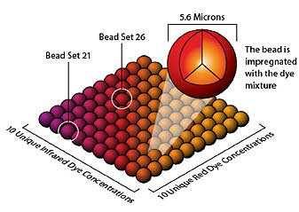
(from Viracor-IBT Laboratory's website)
At the completion of the sandwich immunoassay:
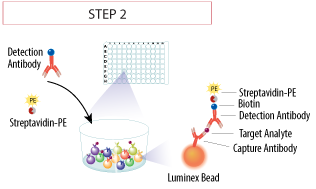
(from R&D Systems website)
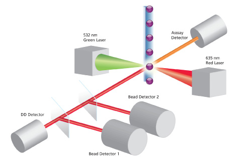
beads are read in single-file by dual lasers for classification and quantification of each analyte using the Bio-Plex Array Reader (Bio-Rad Laboratories Inc., Hercules, CA).
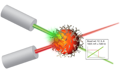
The Bio-Plex array reader is a flow cytometry-based instrument that combines fluidics, 2 lasers, 4 detectors, and real-time digital signal processing to distinguish the 100 different sets of color-coded microspheres, each bearing an analyte-specific assay. The fluidics system of the array reader aligns the microspheres into single file as they enter a stream of sheath fluid and then enter a flow cell:
DartLab Luminex assay request form
Click here to download human multiplex cytokine assay request form.
Click here to download mouse multiplex cytokine assay request form.
DartLab Protocols
Millipore human 41plex measures:
EGF, Eotaxin, FGF-2, Flt-3 ligand, Fractalkine, G-CSF, GM-CSF, GRO, IFN-alpha2, IFN-gamma, IL-10, IL-12 (p40), IL-12 (p70), IL-13, IL-15, IL-17, IL-1ra, IL-1alpha, IL-1beta, IL-2, IL-3, IL-4, IL-5, IL-6, IL-7, IL-8, IL-9, IP-10, MCP-1, MCP-3, MDC (CCL22), MIP-1alpha, MIP-1beta, RANTES, TGFbeta, TNF-alpha, TNF-beta, VEGF, sCD40L
Millipore mouse 32plex measures:
Eotaxin, G-CSF, GM-CSF, IFN-gamma, IL-10, IL-12 (p40), IL-12 (p70), IL-13, IL-15, IL-17, IL-1alpha, IL-1beta, IL-2, IL-2, IL-4, IL-5, IL-6, IL-7, IL-9, IP-10, KC-like, LIF, LIX, M-CSF, MCP-1, MIG, MIP-1alpha, MIP-1beta, MIP-2, RANTES, TNF-alpha, VEGF
Examples
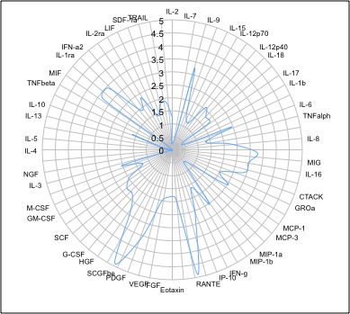
Radar plot showing the concentration of 42 cytokines (pg/ml; log scale) measured in human plasma using a Milliplex kit. Radar plots can be made in Excel: select Charts/Other/Radar.
Methods (for publication)
Cytokines were measured using Millipore human cytokine multiplex kits (EMD Millipore Corporation, Billerica, MA). Calibration curves from reconstituted cytokine standards were prepared with fourfold dilution steps in the same matrix as the samples. High and low controls from the Millipore kits were included. Standards and controls were measured in triplicate; samples were measured once; and blank values were subtracted from all readings. All assays were carried out directly in a 96-well plate (Millipore, Billerica, MA) at room temperature and protected from light.
Briefly, beads together with a standard, sample, control, or blank were added along with their appropriate matrix solution (assay buffer or serum matrix (Millipore, Billerica, MA)) in a final volume of 50 µL and incubated together at room temperature for 2 hours with continuous shaking. Beads were washed three times with 200 µL 1x Wash Buffer (Millipore, Billerica, MA) using a magnetic plate washer. Detection Antibodies (25 µL/well, Millipore, Billerica, MA) were added to the beads for a 1-hour incubation with continuous shaking. No wash was performed after. Then streptavidin-PE (25 µL/well, Millipore, Billerica, MA) was added to the beads for an additional 30-minute incubation. Beads were washed three times with 200 µL 1x Wash Buffer (Millipore, Billerica, MA) using a magnetic plate washer and resuspended in 150 µL of Sheath Fluid (Bio-Rad Cat#171000055) for at least 5 minutes. The fluorescence intensity of the beads was measured using the Bio-Plex array reader. Bio-Plex Manager software with five-parametric curve fitting was used for data analysis.
Links
How to acknowledge Shared Resources
Immunologists Share Top Tips for Multiplex Assay Success
Factors involved in the Cancer Immunity Cycle
Challenges in comparing results to those of traditional methods
Multiplex cytokine assay handbook - includes sample preparation
Bio-Plex magnetic bead assay validation
Luminex extracellular assay protocol (from Invitrogen)
Assay performance of Bio-Plex human multiplex cytokine assay kits
Recent Comments