Spines are prominent postsynaptic morphological features found on the dendrites of many neurons. Many functions for spines have been proposed, including electrical filtering and isolation of synaptic inputs, chemical compartmentalization, and maximization of the number of synaptic connections. However, despite a century of research, a definitive role for dendritic spines remains elusive.
Excitatory postsynaptic potentials (EPSPs) are shaped locally by the dendritic geometry at the site of synaptic input, having larger amplitudes and faster kinetics when generated in neuronal compartments with higher input impedance and smaller local capacitance, as typically occurs at more distal locations in the dendritic tree.As a result, local EPSPs can be highly variable in their amplitudes and kinetics. We hypothesized that spines act to standardize the local postsynaptic geometry experienced by synaptic inputs, limiting location-dependence of local EPSP amplitude and kinetics. To test this hypothesis, we generated computational models of simplified and morphologically realistic dendritic trees and compared the variability of EPSP properties for distributed inputs occurring on dendritic shafts and spines. We found that spines act to standardize the amplitude and kinetics of local EPSPs, limiting their dependence on synapse location within the dendritic tree. This attribute of spines may allow neurons to use similar synaptic mechanisms at all excitatory synapses, regardless of their location within dendritic trees.
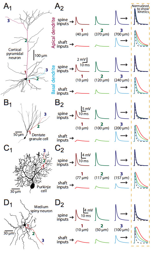
Spines standardize EPSP properties in morphologically realistic neurons. A1-D1: Morphology of reconstructed neurons: a layer 5 pyramidal neuron from the medial prefrontal cortex (A1), a hippocampal dentate granule cell (B1), a Cerebellar Purkinje neuron (C1), and a striatal medium spiny neuron (D1). Synaptic inputs were placed onto shafts and spines of the colored dendrites at proximal, intermediate, and distal locations as indicated by the numbered locations (1 to 3). A2-D2: Left, local EPSPs recorded in spines (top traces) or dendritic shafts (lower traces) at the locations indicated in the different morphologies. Right, normalized and superimposed traces allow comparison of EPSP kinetics. The time course of the underlying synaptic conductance is indicated by a dashed-line.