The cerebral cortex consists of two hemispheres, which are connected to each other by a white matter tract known as the corpus callosum. A specific subset of cortical neurons send axons through the corpus callosum to share information bilaterally within the two cortical hemispheres. Our lab has found that serotonin (5-HT), a ubiquitous neuromodulator in the cortex, selectively activates those cortical neurons that project across the corpus callosum to the contralateral cerebral hemisphere.
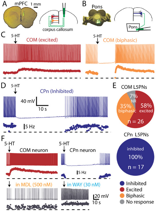
5-HT selectively excites callosal-projection neurons. A: Image of tracer injection location in the prefrontal cortex (left) and schematic of neuron labeling (right). B: Image of trace injection location in the pons (left) and schematic of neuron labeling in the prefrontal cortex(right). C: Excitatory (left) and biphasic (right) responses to focal 5-HT application in callosal projection neurons ("COM" neurons). D: Inhibitory responses to 5-HT in a pontine-projecting ("CPn") neuron. E: Proportion of callosal and pontine projecting neurons excited (red), inhibited (blue), or having biphasic (orange) responses to 5-HT. Non-responsive neurons (NR) shown in grey. F: Serotonergic excitation of callosal projection neurons (left) and inhibition of pontine-projecting neurons (right) was blocked by antagonists specific for 2A (MDL 11939) or 1A (WAY 100635) receptors, respectively.

This figure shows physiological and morphological differences in callosal projecting ("COM") and pontine-projecting ("CPn") neurons. A: Top - Responses to somatic current injections in retrograde-labeled COM and CPn L5PNs. Bottom - Comparison of input resistance (RN) in neurons having different responsiveness to 5-HT. B: Average (± SEM) voltage responses to hyperpolarizing current injections sufficient to generate a peak hyperpolarization of ~20 mV in labeled COM and CPn neurons grouped according to 5-HT response (blue indicates inhibition, red indicates excitation, and orange indicates biphasic responses). Comparison of results is inset. C: Representative morphologies of COM and CPn neurons. D-G: Comparisons of morphological properties in COM and CPn L5PNs (n = 10 for each group). Asterisks indicate p < 0.05.