Our lab has found that transient activation of muscarinic acetylcholine receptors inhibits certain classes of pyramidal neuron in the cerebral cortex and hippocampus. Below are some figures showing the cell-type specificity for the inhibitory effect, and the signal transduction mechanism responsible for the inhibition.
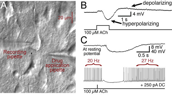
This figure shows a DIC image of a layer 5 pyramidal neuron in the prefrontal cortex (A) and electrical recordings from that neuron (B and C) showing responses to focal ACh application (1 sec duration in B, 40 ms duration in C). Note that ACh hyperpolarizes the pyramidal neuron from resting membrane potentials, and inhibits action potential generation during spiking. A delayed excitation can be seen as depolarization (in B) and increased spike frequency (in C).
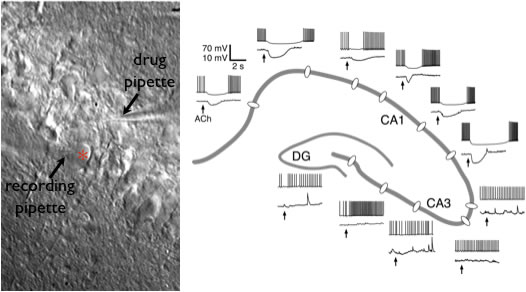
Image showing responses to acetylcholine (ACh) in hippocampal pyramidal neurons. Picture at left is a DIC image of CA1 pyramidal neurons in the hippocampus. The red asterisk indicates the neuron being recorded. A second pipette filled with ACh is placed near the soma. On the right is a map of the hippocampus showing inhibitory responses to ACh in CA1 neurons, but not in CA3 neurons. Lower traces show cholinergic responses at resting membrane potentials, while upper traces are responses during current-evoked action potential generation. ACh also selectively inhibits deep layer neurons in the cerebral cortex, suggesting ACh may have a generalized role in inhibiting cortical output neurons.
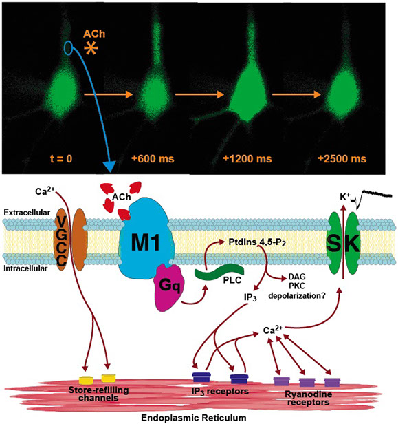
Diagram of inhibitory cholinergic signaling in cortical pyramidal neurons. Top images document a transient increase in intracellular calcium occurring after a brief application (20 ms) of acetylcholine near the apical dendrite of a layer 5 pyramidal neuron (asterisk at time 0). The calcium transient begins in the apical dendrite several hundred milliseconds after ACh application, then spreads to the soma and basal dendrites. Peak calcium occurs about 1200 ms following ACh application, and the event is over by 2500 ms. Diagram below shows the intracellular signaling cascade that acetylcholine initiates to produce calcium increases and inhibition of the pyramidal neuron.