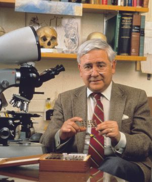
Returning from a recent trip to his native Spain, 89-year-old Emeritus Professor Miguel Marin-Padilla made a surprising decision—after nearly 60 years in academic medicine and more than 190 scientific publications, it was time to leave science behind. “It is time for me to close that door and let young people move the field forward,” he says.
Marin-Padilla was invited to his alma mater in Granada to accept an honor membership in the Spanish Society of Anatomical Pathology and the International Academy of Pathology—in recognition of his career and body of work dedicated to the field of pathology. While there, he also visited the Institute of Neurosciences of Castilla y León in Salamanca where he met with enthusiastic graduate students and gave a talk on the repair of perinatal brain damage in the pathogenesis of epilepsy.
An emeritus professor of pathology and of pediatrics, Marin-Padilla has had a long and fulfilling career. For more than 40 years, he taught general pathology to Dartmouth medical students, and developmental pathology to pediatricians and neonatologists at Dartmouth-Hitchcock.
In the late 1970s he became interested in studying the brain and has since devoted his life to that pursuit. His research garnered the Jacob Javits Neuroscience Investigator Award—given by the National Institutes of Health’s National Institute of Neurological Disorders and Stroke to investigators of “exceptional talent, imagination, and preeminent scientific achievement.”
And his retirement, 18 years ago, did little to stop his quest to understand the brain. Marin-Padilla has spent nearly all of those years conducting research in his "Sancta Sanctorum," which houses his collection of more than 5,000 pristine rapid Golgi preparations (a method of staining nerve tissue allowing neurons to be seen with great clarity), an old microscope, and books with drawings by Santiago Ramón y Cajal.
Ramón y Cajal, a Spanish neuroscientist and pathologist, who in the late 1880s improved Camillo Golgi’s staining method to study neurons, found that the brain’s nervous system is made up of individual cells touching one another, rather than a network—proving for the first time that neurons behave as biochemically distinct cells—a discovery that became the basis of modern neuroscience. Both Golgi and Ramón y Cajal made beautiful drawings of brain cells they saw in microscopic bits of brain tissue.
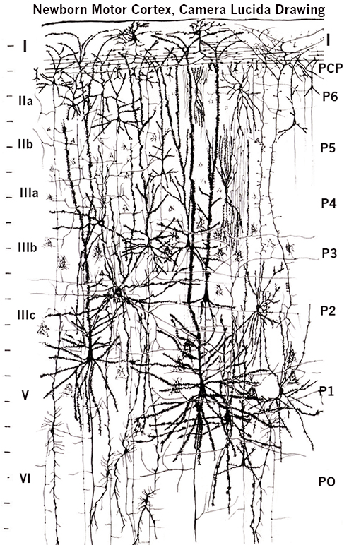
Captivated by Ramón y Cajal’s work, Marin-Padilla has used the same centuries-old methods for more than 50 years—cutting brain tissue into tiny sections by hand with a razor blade, staining and preparing them for study, which requires painstaking patience and dedication—to map the activity of neurons in the human brain. Setting him apart from other neuroscientists, he too made artistic and elegant drawings illustrating what he saw through the microscope.
But few 21st century neuroscientists are using Golgi’s method. They use modern techniques, such as computer modeling and crowd sourcing to answer fundamental questions about the brain.
His dedication to the 19th century technique has led to many important discoveries: he was one of the first to describe pioneer neurons that are critical in setting up the pathways by which all other neurons in the brain migrate to their proper place; he dispelled a common belief that neurons extend their branches up to the superficial layers of the cortex—they elongate from the top of the brain downward; was the first to link structural abnormalities in the brain—that abnormally shaped neurons correlated with cognitive difficulties in children with Down syndrome and those who developed epilepsy as a result of perinatal brain damage; he provided detailed descriptions of how blood vessels grow into the brain as it develops; and contributed to understanding the fundamental principles that govern the embryonic origins of the mammalian cerebral cortex.
On June 3, Marin-Padilla published what he says is his final paper—“Endothelial Cells Filopodia in the Anastomosis of Central Nervous System Capillaries” in Frontiers in Neuroanatomy—capping a career of more than 190 publications focusing mostly on the motor cortex, including two books.
But Marin-Padilla has no intention of pursing a life of leisure. “I cannot stay put, I have to keep going,” says the lifelong walker who still logs two miles each day mulling over new questions and ideas—though his thoughts no longer compel him to return to the Sancta Sanctorum and his brain slides.
“I know a great deal about the human brain—I’ve spent a lifetime studying it, so I’m going to write novels based on my understanding,” he says smiling. “One of my ideas takes place in prehistoric times, it’s about what happens to identical twins when one of them suffers a brain injury and develops a new personality.”
He hasn’t completely left science behind.