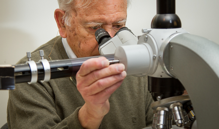
In the late 19th century, the Italian scientist Camillo Golgi developed a painstaking staining technique that revealed fine details of the nervous system. Continuing in that tradition, the Geisel School of Medicine’s Miguel Marin-Padilla has been using Golgi’s method to unravel the brain’s secrets for nearly five decades.
By permeating brain tissue with potassium dichromate and silver nitrate throughout a four-day process, the method clearly stains a random selection of cells in their entirety and reveals the connections between neurons. In the late 19th and early 20th centuries, Spanish scientist Santiago Ramón y Cajal adapted and used the technique in his pioneering investigations of the microscopic structure of the brain, and, in 1906, Cajal and Golgi shared the Nobel Prize in Physiology or Medicine for their work. To find out how Cajal, considered by many to be the father of modern neuroscience, was able to see and understand so much, Marin-Padilla traveled to the Cajal Institute in Madrid, Spain, in the late 1960s, when he was starting his research career at Dartmouth, on a National Institutes of Health Neurohistology Fellowship to study the technique.
Learning Golgi’s method profoundly influenced his career as a researcher, “I also learned how patient you must be,” Marin-Padilla recalls. “I brought that knowledge back to Hanover and started using it. Everyone thought I was crazy for using a method so old. But I knew it was a good method, and I had plenty of time during the long New England winters to use it. I did everything myself—as director of pediatric pathology I obtained and cut the brain by hand with a razor blade into small sections, stained and prepared them, and began to study.”
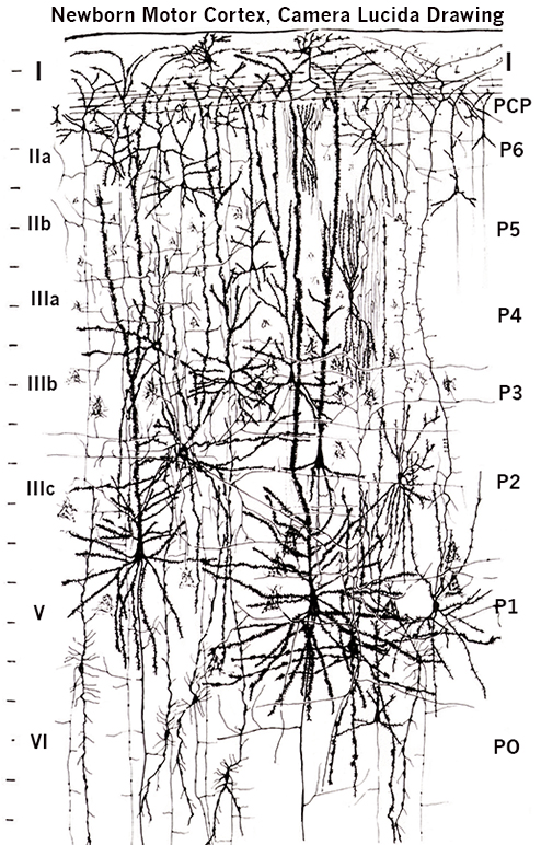
Following in Golgi’s and Cajal’s footsteps—both made beautiful drawings of what they saw through their microscopes—Marin-Padilla created artistic and elegant drawings that illustrate what he sees in thousands of miniscule bits of brain tissue. His dedication to the Golgi method has led to many important discoveries.
His work was instrumental in establishing that neurons do not extend their branches up to the superficial layers of the cortex (as was commonly believed), but elongate from the top of the brain downward. He provided some of the first information to show that abnormally shaped neurons correlated with cognitive difficulties in children with Down syndrome or those who developed epilepsy as a result of perinatal brain damage. He has provided detailed descriptions of how blood vessels grow into the brain as it develops. Finally, his work has contributed to understanding the fundamental principles that govern the embryonic origins of the mammalian cerebral cortex.
“These discoveries opened a big window into brain studies because now we know that neurological disorders may be related to structurally abnormal neurons rather than to abnormal brain neurochemistry,” Marin-Padilla says. “All of a sudden there is a huge field in front of us to be investigated.”
“He’s an amazing individual,” says Leslie Henderson, a professor of physiology and neurobiology at Geisel, who has known Marin-Padilla for 25 years. “He is someone who has made very fundamental and insightful discoveries that have changed neuroscience. He was one of the first to describe pioneer neurons, and as their name implies, they are critical in setting up the pathways by which all other neurons in the brain migrate to their proper place—it was a major and fundamental discovery.”
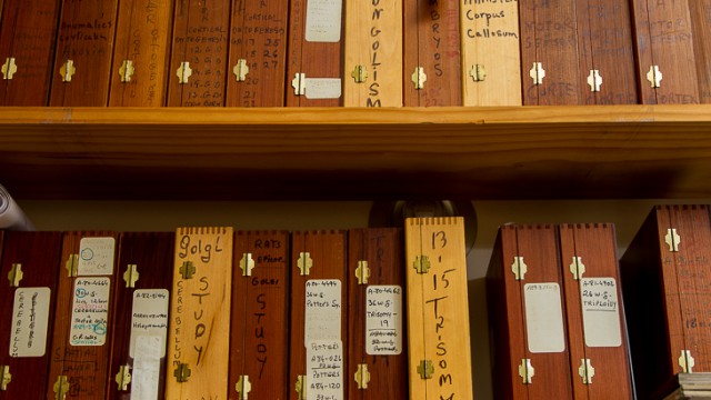
Since his retirement 14 years ago, Marin-Padilla has spent a fair amount of time in a room at his house that he calls the "Sancta Sanctorum," which houses his collection of more than 5,000 pristine rapid Golgi preparations, his old microscope, and many books.
He has contributed to several books on neuroscience and has gathered his Golgi observations into a book on the prenatal development and structure of the human brain motor cortex—The Human Brain: Prenatal Development and Structure (Springer, 2011), which is dedicated to his beloved late wife, Teresa.
“When I get an idea,” he says. “I go down to my Sancta Sanctorum, and to my brain slides, to see if the idea means something, to see if I’m right or if I’m wrong.”
His recent paper, his 158th, on the brain’s vascular system, “The Human Brain Intracerebral Microvascular System: Development, Structure and Function,” published this winter in Frontiers in Neuroanatomy, has captured the imagination and interest of scientists.
“Miguel has been publishing since the 1950s and not just in obscure journals,” Henderson says. “It’s phenomenal that he’s had this type of longevity in a scientific career to still be writing about things that people are interested in reading.”
Exploring the Human Brain
In 1977, Marin-Padilla wrote an article for the medical school's alumni magazine about how he first became interested in studying the brain. Read the full article here [PDF].
While Marin-Padilla is humble about his accomplishments, he is aware that the technique he believes in and has used for nearly 50 years sets him apart from other neuroscientists. And because of the patience and dedication the Golgi method requires, there are few, if any, willing to follow in his footsteps.
“There is nobody doing this type of work now in the same way that Miguel does,” Henderson says. “The BRAIN Initiative is, in essence, doing the same thing Miguel has tried to do on his own—map the activity of every neuron in the human brain—but using slightly different techniques, 21st-century techniques such as crowd sourcing, to answer fundamental questions about neurons. What Miguel has devoted his life to has now moved into the social media space.”
Enthusiastic and as devoted as ever to using the Golgi method, Marin-Padilla isn’t sure what the future holds.
“But I do know one thing,” he says. “I will continue to study the human brain neuronal, vascular, and glial structure—I don’t know what I’m going to find along the way. What’s amazing to me is that I’m going to be 84 years old in July and I’m still pursuing this dream!”
Dr. Marin-Padilla taught general pathology to Dartmouth medical students and developmental pathology to pediatricians and neonatologists for more than 40 years. At the Geisel School of Medicine, the Miguel Marin-Padilla Lectureship for Excellence in Medical Education and the Miguel Marin-Padilla Medal were established in his honor.